 400-029-0925
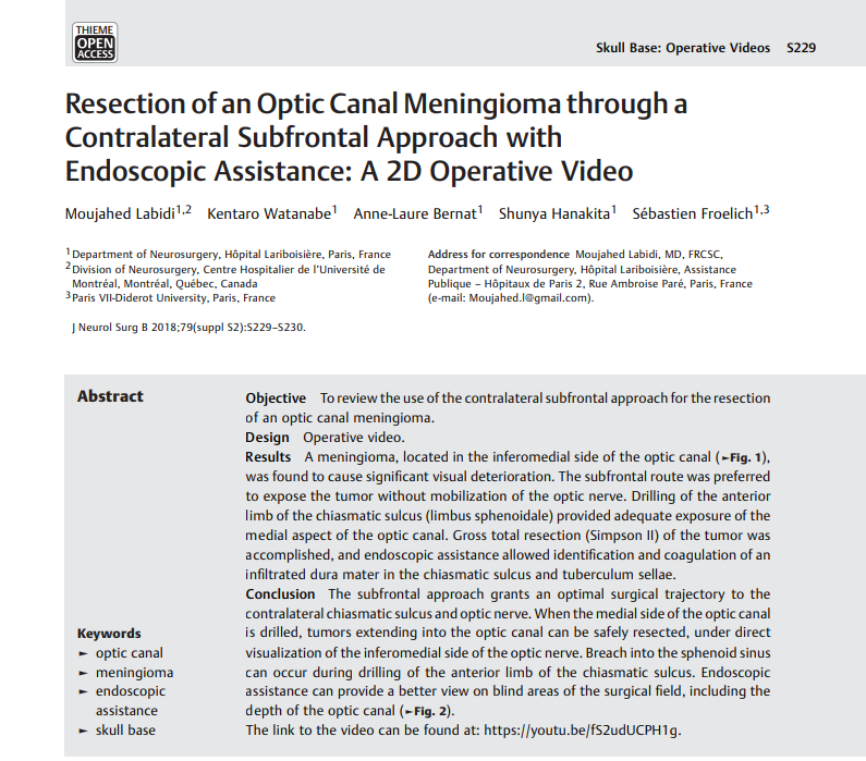
Resection of an Optic Canal Meningioma through a Contralateral Subfrontal Approach with Endoscopic Assistance: A 2D Operative Video
内镜辅助下对侧额下入路切除视神经管脑膜瘤:二维手术视频
英文摘要:
Objective To review the use of the contralateral subfrontal approach for the resection of an optic canal meningioma.
Design: Operative video.
Results A meningioma, located in the inferomedial side of the optic canal ,was found to cause significant visual deterioration. The subfrontal route was preferred to expose the tumor without mobilization of the optic nerve. Drilling of the anterior limb of the chiasmatic sulcus (limbus sphenoidale) provided adequate exposure of the medial aspect of the optic canal. Gross total resection (Simpson II) of the tumor was accomplished, and endoscopic assistance allowed identification and coagulation of an infiltrated dura mater in the chiasmatic sulcus and tuberculum sellae.
Conclusion The subfrontal approach grants an optimal surgical trajectory to the contralateral chiasmatic sulcus and optic nerve. When the medial side of the optic canal is drilled, tumors extending into the optic canal can be safely resected, under direct visualization of the inferomedial side of the optic nerve. Breach into the sphenoid sinus can occur during drilling of the anterior limb of the chiasmatic sulcus. Endoscopic assistance can provide a better view on blind areas of the surgical field, including the depth of the optic canal.
The link to the video can be found at: https://youtu.be/fS2udUCPH1g.
中文摘要:
目的:探讨对侧额下入路切除视神经管脑膜瘤的手术方法。
设计:手术视频。
结果:脑膜瘤,位于视神经管下内侧,被发现会导致明显的视力下降。优选额下路径暴露肿瘤而不动视神经。交叉沟(蝶缘)前肢的钻孔提供了足够的视神经管内侧的暴露。完成了肿瘤的全部切除(Simpson II),内镜辅助下可在交叉沟和鞍结节处发现并凝固浸润的硬脑膜。
结论:额下入路为对侧交叉沟及视神经提供了更佳的手术路径。当钻取视神经管内侧时,在视神经下内侧的直接可视化下,可以安全地切除伸入视神经管的肿瘤。在交叉沟前肢钻孔时,可发生蝶窦破裂。内镜辅助可以更好地观察手术领域的盲区,包括视神经管的深度。
该视频的链接可以在https://youtu.be/fS2udUCPH1g找到。
INC国际神经外科医生集团旗下组织世界神经外科顾问团(WANG)成员,世界神经外联合会(WFNS)颅底手术委员会主席(2013年至今)Sebastien Froelich教授,是世界知名的神经外科内镜手术专家,他对于脑膜瘤、脊索瘤、垂体瘤、颅咽管瘤等都有大量的临床治疗经验。Sebastien Froelich教授尤其擅长神经内镜鼻内入路的颅底肿瘤切除,针对脑膜瘤、垂体瘤、脊索瘤、等采取神经内镜下的微创手术。其发明的知名的内镜手术“筷子手法”操作方式不止提高了肿瘤的切除率,更是使肿瘤患者有了更好的预后效果。
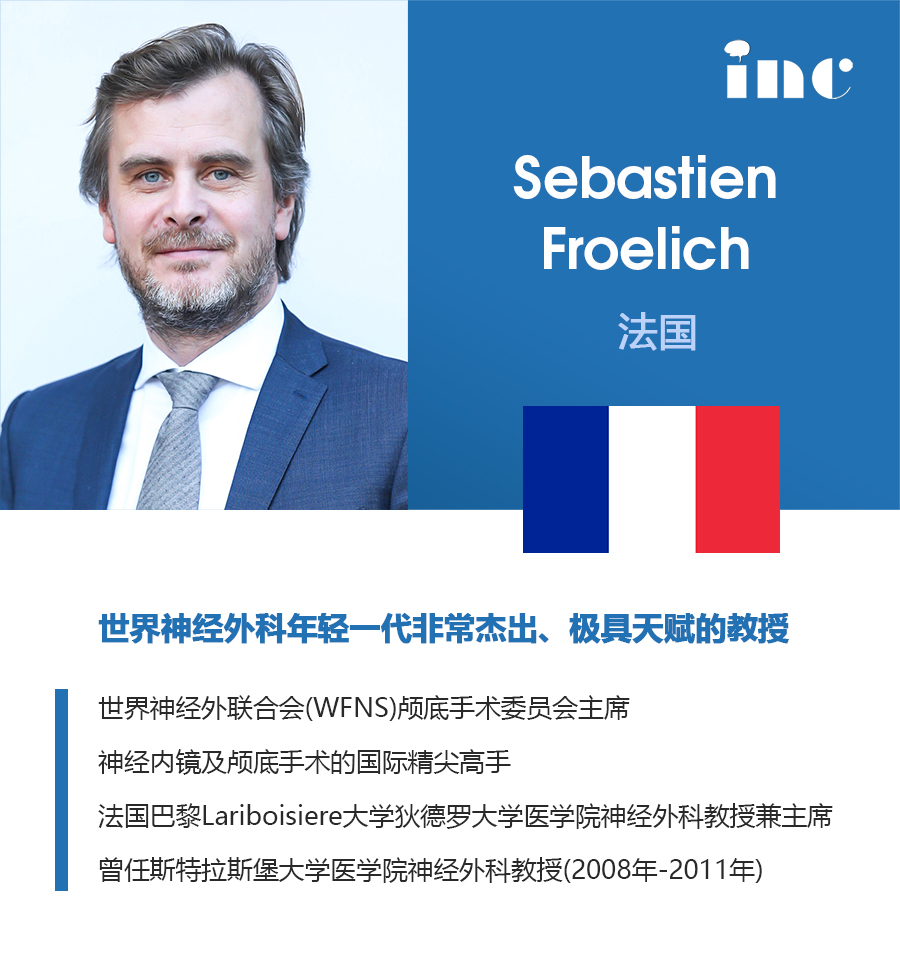
9月7日、8日,世界神经外科联合会(WFNS)2019全球大会的多个会前会在首都医科大学宣武医院召开,Sebastien Froelich教授在宣武医院开展现场指导教学,内容涵盖手术展示及Storz脑室镜模拟训练、复杂颅底入路的解剖等,学员们纷纷表示收获颇丰。9月10日,Sebastien Froelich教授在WFNS大会上发表了《颅颈交界区肿瘤的治疗》主题演讲,以大量的临床案例和手术视频介绍了神经内镜技术如何对各类颅经交界处肿瘤进行精准切除,并系统总结了神经内镜技术操作要点和手法技巧等。Froelich教授高超的内镜下手法和显微外科手术技巧都让参会者叹为观止。Sebastien Froelich教授为中国神经内镜技术的发展起到了一个指导性的作用。
INC国际神经外科医生集团旗下组织世界神经外科顾问团(WANG)有多位世界闻名的神经外科教授,他们分别代表美国、欧洲、日本乃至全世界至高的神经外科水平,分别任职各自领域的世界相关协会主席。INC国际一直致力于中外神经外科技术的交流、合作、促进和提高,同时针对中国有需要的脑膜瘤、脊索瘤、听神经瘤、垂体瘤、颅咽管瘤、脑胶质瘤、脊髓肿瘤、脑血管畸形病变、动脉瘤等神经外科高端患者,提供国际治疗咨询与协调服务。
- 文章标题:内镜辅助下对侧额下入路切除视神经管脑膜瘤:二维手术视频
- 更新时间:2019-09-16 16:20:27
INC国际神经外科医生集团,国内脑瘤患者治疗新选择,足不出户听取世界神经外科大咖前沿诊疗意见不是梦。关注“INC国际神经科学”微信公众号查看脑瘤治疗前沿资讯,健康咨询热线400-029-0925,点击立即预约或在线咨询直接预约INC国际教授远程咨询!