 400-029-0925
Microelectrode-guided posteroventral pallidotomy for treatment of Parkinson's disease: Postoperative magnetic resonance imaging analysis(微电较引导后腹苍白球切开术治疗帕金森氏病:术后磁共振成像分析)
英文原文摘要
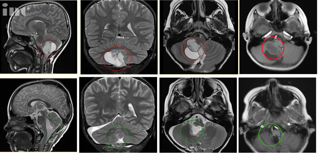
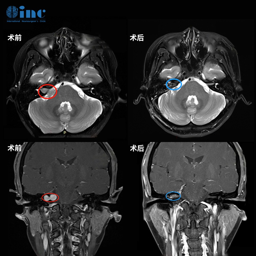
The authors report the postoperative magnetic resonance (MR) imaging findings in 36 patients with advanced Parkinson's disease who underwent unilateral microelectrode-guided posteroventral pallidotomy. The lesions were placed within 1 mm of the ventral border of the globus pallidus internus (GPi) to include pallidothalamic outflow pathways. Sequential MR studies were obtained within 1 to 3 days postoperatively and at 6-month follow-up examination. Thirty-four (94%) of the 36 patients enjoyed sustained moderate or marked improvement of their parkinsonian symptoms 6 months postoperatively. Transient side effects occurred in five patients (14%), but there were no persistent complications. The pallidal radiofrequency lesions were prolate spheroid shaped and were composed of three concentric zones in the early postoperative studies. The mean volume of the middle zone, corresponding to the area of hemorrhagic coagulation necrosis, was 44.4 ± 17.6 mm3; the mean lesion volume as defined by the outer zone, corresponding to perilesional edema, was 262.2 ± 111.6 mm3. Additional edema spreading to the internal capsule was noted in 32 of 34 cases and to the optic tract in 11 of 34 cases. In two patients small ischemic infarctions involving the corona radiata were found, and in one a venous infarction was detected. Ischemic infarction resulted in mild transient Broca's aphasia in one patient, but there was no detectable neurological deficit in the other two. The mean volume of late-phase (6 months) lesions was 22 ± 28.8 mm3. In three patients no lesion was identified despite sustained clinical improvement. The lesion was located in the posteroventral GPi in all cases except in one patient in whom it was confined to the GP externus (GPe). This 49-year-old woman did not experience sustained benefit. The authors found no consistent correlations between lesion size and location and clinical outcome as measured by a global outcome score, the Unified Parkinson's Disease Rating Scale motor, activities of daily living, and bradykinesia 'off' scores or rating of dyskinesias. Lesioning of pallidal and subpallidal pathways may contribute to the sustained clinical benefit in this series. Magnetic resonance imaging analysis showed that intra-operative microelectrode recording facilitated accurate placement of the lesion in this critical area.
中文摘要
作者报告了36例接受单侧微电较引导的后腹苍白球切除术的晚期帕金森氏病患者的磁共振成像结果。将病变置于距苍白球内侧(GPi)腹侧边界1 mm以内,包括苍白丘脑流出途径。术后1到3天内和6个月的随访检查中进行了顺序MR研究。术后6个月,36例患者中有34例(94%)的帕金森病症状持续持续好转。五名患者(14%)发生了短暂的副作用,但没有持续的并发症。在术后早期研究中,苍白的射频损伤呈扁球形,并由三个同心区组成。由外部区域确定的平均病灶体积对应于病灶周围水肿,为262.2±111.6 mm。在34例中有32例中发现有其他水肿扩散到内囊,在34例中有11例又扩散到视道。在两名患者中发现了涉及电晕放射线的小型缺血性梗塞,在一名患者中发现了静脉梗塞。缺血性梗塞导致一名患者出现轻度短暂性Broca失语症,但另一名患者未发现可检测到的神经功能缺损。晚期(6个月)病变的平均体积为22±28.8 mm。尽管有持续的临床好转,但在三名患者中未发现病变。在全部情况下,病变均位于后腹GPi,只有一名患者局限于GP外部(GPe)。这名49岁妇女没有持续受益。作者发现,通过总体结果评分,统一帕金森氏疾病评分量表运动,日常生活活动以及运动迟缓“关闭”评分或运动障碍评分,病变大小和位置与临床结果之间没有一致的相关性。苍白和苍白下途径的病变可能有助于该系列的持续临床获益。磁共振成像分析表明,术中微电较记录促进了病变在该关键区域的准确放置。
该论文的一作者为Joachim K. Krauss教授,他目前是国际立体定向与功能神经外科学会主席、欧洲立体定向与功能神经外科学现任荣誉主席、世界神经外科联合会(WFNS)立体定向与功能神经外科委员会主席,而且也是INC国际神经外科医生集团旗下世界神经外科顾问团成员。
Joachim K. Krauss教授的擅长包括神经肿瘤学、小儿神经外科、血管神经外科、颅底神经外科、脊柱外科、脊柱外科、重建立体定向与功能神经外科、创伤神经外科、疼痛神经外科、脑积水外科等,涵盖面较为广泛,是神经外科领域的全能型专家。与此同时,Joachim K. Krauss教授在汉诺威医学院(MHH)中的医疗团队由高度化的专家组成,并在神经外科治疗中合适使用计算机断层扫描(CT)和磁共振成像(MRI),从而合适将患者的治疗提高到平均值以上。
1990年代初,Jean Siegfried在苏黎世创立了深部的脑电较刺激DBS手术疗法,作为治疗帕金森氏症的替代疗法。然而在后续的运用中,Joachim K. Krauss教授和法国的Coubes教授在上世纪90年代末将其好转,并具有提出性的运用在治疗肌张力障碍上。最初,Joachim K. Krauss教授的方式遭到了医学界的质疑,然而在随后的多次研究中,DBS手术疗法被证实在治疗肌张力障碍上发挥了重要的作用,可作为肌张力障碍患者选择的靶点之一。教授也因此贡献在2002年获得德国肌张力障碍奥本海姆奖。
当然,Joachim K. Krauss教授除了在肌张力障碍的治疗上做出了贡献之外,在基地神经节神经药理学方面也获得了不小的成就,并由此获得了博士学位。在工作期间,Joachim K. Krauss教授曾在德国海德堡大学做过副教授,并在美国德克萨斯州休斯顿的贝勒医学院担任过兼职副教授。随后通过自身优异的表现,2001年在国际立体定向和功能神经外科协会担任主任一职,并于2005年被国际立体定向和功能神经外科学会任命为主席。Joachim K. Krauss教授目前在汉诺威医学院担任主任,并在神经外科学系任教。
- 文章标题:微电较引导后腹苍白球切开术治疗帕金森氏病:术后磁共振成像分析
- 更新时间:2019-10-31 16:50:08